AMD VR
Project
General Problem
Macular Degeneration is a deterioration of the central portion of the retina. The macular is part of the retina in the inner eye and contains a high density of photoreceptor cells. If this part deteriorates a growing blind spot forms that significantly affects vision. Nerve cells are dying off in the macula. Once dead, these cells cannot be repaired. Macular Degeneration is a leading cause of vision loss in the USA. It can be caused by multiple reasons e.g. related to smoking, or genetics, but is very often age related. Age-related Macular Degeneration (AMD) is the leading cause of vision loss among persons over 50 years.
Project Approach
We designed and prototyped a VR-based assistive technology for patients in a collaboration between Georgia Tech and Emory University. The project focuses on two separate deliverables: 1) a VR-based interface for patients that optimizes their field of view for their particular AMD condition 2) a desktop interface for experts to assess and optimize the digital mapping for such a VR visualization.
The system uses commercially available VR equipment (HTC Vive Pro Eye) to build an experimental set up investigating digital media opportunities for people affected by Macular Degeneration. Its initial user scenario targets home media use. The design aims at supporting reading, writing, watching TV, and computer interactions. Currently these are supported by special magnifying technologies. Based on earlier work (Juday/ Loshin 1988) our approach warps the image through interactive mapping in the VR visualization to optimize it for each eye’s remaining active retina. This differs in its design from magnification alone and potentially offers a new venue in this area. It has been shown that vision warping is eventually corrected by the brain to create a produce a complete picture of the world around.
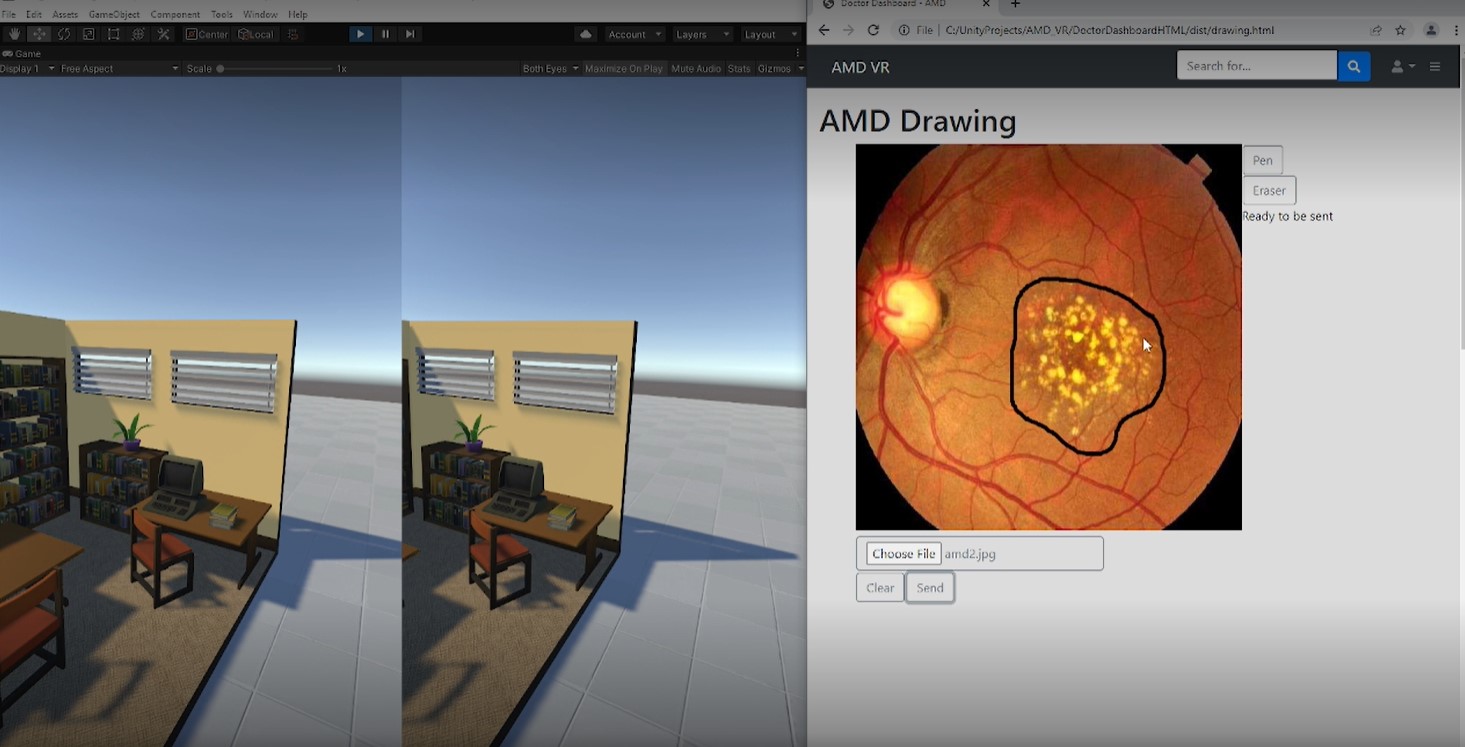
The second application is the necessary interface for the doctor, which allows eye care specialists to assess the patient’s condition and adjust the digitally enhanced field optimization. Doctors can activate visions charts, change contrast, hand-adjust the occluded area, and activate the warping effect while the patient experiences these changes in real-time in the VR viewpoint. This allows the necessary adjustment of the digital image and provides the expert with the tools needed to make this kind of VR enhanced visualization useful for patients. The design allows doctors to adjust the conditions on each eye separately to optimize the visualization for a particular patient’s situation. For example, the size, shape, or position of the warping effect can be manipulated for each eye individually.
Existing Work
Existing AMD treatments largely target a slowing down of the deterioration. They include in-eye-injections, nutritional supplements (AREDS). Depending on the particular case, other treatment consists of laser surgery or photodynamic therapy – both techniques are invasive and can have adversarial side effects. There are additional, still experimental, surgical approaches. There is no cure or reversal of this process. Since 2010 the FDA has approved special implantable miniature telescope (IMT) prosthetic devices into eyes. There are different techniques depending on the implant, but basically the IMT implant sits in the eye and adjusts the image before it hits the macula. Most supportive non-invasive methods consist of devices such as specialized glasses and other magnifying technologies. The closest related work to the here proposed work are digitally enhanced magnifying devices.
See, for example:
Jordy or eSight or NuEyes